STEP ONE

Be informed.  Read information below.

STEP TWO

Schedule consultation with R. Douglas Wichman, MD.

STEP THREE

Submit "New Patient Form", current labwork, & imaging results prior to consultation.

STEP FOUR

Establish if you are a candidate for this therapy

in accordance with the Georgia

"Access to Medical Treatment Act".


Image result for oncolytic virus


----------------------------------------------------------------------------------------------------------------------------------------------------------

*** SPECIAL NOTE FROM DR. WICHMAN ***

The following excellent article was reproduced from Melanoma Research at https://www.ncbi.nlm.nih.gov/pmc/articles/PMC4560272/:


Melanoma Res. 2015 Oct; 25(5): 421–426.
Published online 2015 Sep 3. doi:  10.1097/CMR.0000000000000180
PMCID: PMC4560272

Adapted ECHO-7 virus Rigvir immunotherapy (oncolytic virotherapy) prolongs survival in melanoma patients after surgical excision of the tumour in a retrospective study


Introduction

Melanoma is one of the fastest-growing cancers and has the highest mortality rate of skin cancers 13. More than half of melanoma patients experience progression of the disease within 3 years of diagnosis 4,5. Current clinical practice guidelines for stage I–II melanoma provide few, if any, recommendations for treatment 69. The oncolytic property of viruses has been observed for over a century and is presently being studied intensively 1016. An oncolytic, nonpathogenic ECHO-7 virus, adapted and selected for melanoma that has not been genetically modified (Rigvir), was approved and registered in 2004 in Latvia for melanoma therapy 1727. The effect of viruses on cancers, including melanoma, has been tested in clinical trials; however, the effectiveness of an approved and marketed virus has not yet been shown in a clinical setting 12,14,16.

In oncolytic virotherapy, Rigvir is a first-in-class. At a later time, a genetically modified adenovirus was approved for head and neck cancer 28,29. Melanoma is staged with substages from 0 to IV by measuring the thickness of the tumour according to Breslow, by assessment of ulceration, mitotic rate and metastases and by collecting pathologic information on regional lymph nodes 30,31. On the basis of the stage of the disease, treatment is currently performed according to published guidelines 4,69.

The aim of the present study is to test the effectiveness of Rigvir in a retrospective study in substage IB, IIA, IIB and IIC melanoma patients on time to progression and overall survival.

Methods

Retrospective clinical study patients

White patients (N=79) who had undergone surgical excision of melanoma and diagnosis verified histologically during the 4 years between January 2008 and December 2011 were included in this study. All patients were free of disease after surgery and were classified into substages IB, IIA, IIB and IIC according to the American Joint Committee on Cancer 30,31. For disease progression, all were followed for a minimum of 3 months until January 2014. The overall survival was checked on 5 June 2014 and considered to reflect the status by 27 May 2014. The detailed study population characteristics of this retrospective study are shown in Table Table11.

Table 1
Study population characteristics (N=79)

Current guidelines for melanoma advise no treatment postsurgery for patients who are classified into substages IB and IIA. Patients in substages IIB and IIC are provided three options: participation in a clinical trial, observation and interferon 7,8. In the absence of strict guidelines, treatment with Rigvir was offered. Thus, 52 study participants received Rigvir and 27 were observed according to the guidelines. The patients who had been treated with interferon were excluded from the present analysis as, in the registry, they were too few to allow for any comparison.

As a part of the safety assessment, serum clinical chemistry parameters were recorded.

The patients in this study were treated in the Latvian Oncology Center of Riga Eastern Clinical University Hospital, the Latvian Virotherapy Center in Riga and the Oncology Clinic of Piejūras Hospital in Liepāja, Latvia.

The study was approved by the respective ethics committee.

Rigvir characteristics

Rigvir is a 2 ml frozen solution of an adapted and selected ECHO-7 virus strain, Picornaviridae family, Enterovirus genus, Enteric Cytopathic Human Orphan (ECHO) type 7, group IV, positive-sense single-stranded RNA virus produced under GMP. The titre is not less than 106 TCID50/ml in sodium chloride for injection.

Method of Rigvir administration

Treatment was started after surgical excision of the primary melanoma tumour when the wound had healed. First, Rigvir (2 ml) was administered intramuscularly regionally for 3 consecutive days. After about 4 weeks, administration was repeated for three consecutive days and repeated about 4 weeks later. Subsequently, a single administration of Rigvir (2 ml, intramuscularly) was performed at monthly intervals during the first year, at 6-week intervals during the first half of the second year, at 2-month intervals during the second half of the second year and at 3-month intervals in the third year. Rigvir is not to be used during an acute infection.

Statistical analysis

Statistical analysis of the data was carried out using the SPSS statistical software, V.20 (SPSS Inc., Chicago, Illinois, USA). Mann–Whitney U-test and Wilcoxon tests (for continuous variables), Fisher’s exact test and the χ2-test (for categorical variables) were used to test differences between and within groups. Cox proportional hazard survival regression analysis was carried out, which is the most commonly used multivariate model in survival analysis. Thus, any difference between the groups, for example, in age, has been taken into account in the Cox analysis. (This is in contrast to Kaplan–Meier analysis, which is a bivariate analysis that only takes into account one predictor at a time). Hazard ratios (HRs) and 95% confidence intervals were calculated using bivariate and multivariate Cox regression analysis on survival. Endpoints were occurrence of metastases or disease recurrence for time to progression, and death from any cause for analysis of overall survival. Predictors (covariates) used in regression analysis were tumour stages, treatment (Rigvir, observation), sex and age. A P value less than 0.05 from a two-sided test was established to indicate statistical significance.

Results

Effectiveness in patients: time to progression

Melanoma patients of substages IB, IIA, IIB and IIC were studied according to the postsurgery management that they had received. One group was treated with Rigvir and the other was managed according to current guidelines by observation (the control group is called ‘observation’) 69. The follow-up period was not statistically different between both treatment groups (Table (Table11).

Patients who were free of melanoma after surgical excision and were treated with Rigvir appeared to remain disease free (free of metastases and/or recurrence) for a longer period of time compared with a similar group of patients who did not receive Rigvir. The difference between the treatment groups did not, however, reach statistical significance (Table (Table22).

Table 2
Regression estimates from Cox regression analysis of time to progression (N=79)

Effectiveness in patients: overall survival

The survival of patients who were treated with Rigvir was significantly (P<0.05) longer compared with a similar group of patients who did not receive Rigvir (Fig. (Fig.11 and Table Table3).3). The difference between both treatment groups was statistically significant on analysing all four substages together (IB, IIA, IIB, IIC) (Table (Table3)3) and on analysing stage II together (substages IIA, IIB, IIC). Adjusting for patient age, sex and substage of disease, the HR was calculated in multivariate Cox regression analysis. The HR for patients treated according to current guidelines by observation versus treated with Rigvir was 6.27 (P<0.005) for all patients, 4.39 (P<0.032) for substage IIA–IIC patients and 6.57 (P<0.014) for substage IIB–IIC patients (Fig. (Fig.1).1). This indicates that the patients who were treated with Rigvir had a 4.39–6.57-fold lower mortality than those treated using current guidelines by observation.

Fig. 1
Cox regression analysis plots of survival of melanoma patients following surgery. P is the statistical significance of the difference between the Rigvir (—) group and the observation according to current guidelines (observation) group (---) after ...
Table 3
Regression estimates from Cox regression analysis of survival (N=79)

Safety assessment

In the previous clinical studies, a few side effects were reported, for example subfebrile temperature (37.5°C for a couple of days), pain in the tumour area, sleepiness and diarrhoea. In this retrospective study, however, there was no record of any untoward side effect from Rigvir treatment or its discontinuation.

Serum clinical chemistry parameters were recorded and graded according to NCI CTCAE 32 (Table (Table4).4). In the observation group, grade 1–3 values were obtained. All grade 3 samples were from two patients obtained within the last few months of life. In one of these patients, progression of the disease was reported simultaneously. In contrast, in the Rigvir-treated patients, values above grade 2 were not observed.

Table 4
Levels of serum clinical chemistry parameters during treatment

Discussion

Oncolytic virotherapy is one of three forms of virotherapy (the other two being viral vectors for gene therapy and viral immunotherapy, respectively). Early observations of tumour regressions after virus infections have been published starting from the late 19th century (cf. 1016). Recently, several oncolytic viruses have been tested clinically 3335 and Science named cancer immunotherapy the breakthrough of the year of 2013 36. The melanoma adapted and selected ECHO-7 virus Rigvir is first-in-class in oncolytic virotherapy; it is approved as therapy for melanoma.

The present results show that in substage IB, IIA, IIB and IIC melanoma patients, Rigvir administration after surgery significantly (P<0.05) prolongs survival compared with patients who were managed according to current published guidelines 69. For the Rigvir-treated patients, the HR (risk of death) is 4.39–6.57-fold lower than for the control group treated according to current guidelines by observation. The HR was calculated in multivariate Cox regression analysis adjusting for patient age, sex and substage of disease.

In this study, there was no record of any untoward side effect from Rigvir treatment, which is in agreement with clinical studies using other oncolytic viruses 14,16,33,34,37. Moreover, no value higher than grade 2 was recorded in Rigvir-treated patients. This is in contrast to most other cancer therapies, where grades 3 and 4 are frequently observed (cf. 38).

Administration of virus induces the formation of neutralising antibodies that might potentially influence the efficiency of Rigvir. In previous studies, the titre of neutralising antibodies against ECHO-7 was determined in both healthy individuals and patients before administration of Rigvir. In 94 healthy adult participants tested, the titres were found to be low (1 : 20 to 1 : 62) 39,40. When tested in 155 adult cancer patients who had not been treated with Rigvir, neutralising antibodies against ECHO-7 were detected in ∼50% of the patients 41. In a local study of 472 individuals, the presence of ECHO-7 antibodies was shown to increase with age in children and level off to a plateau of around 75% in adults 42. To our knowledge, the prevalence of neutralising antibodies against the ECHO-7 virus in the general adult population has not been reported.

Rigvir is an immunomodulator that affects both the humoral, antibody-mediated, and the cellular immune systems 2022. When virus adsorption and penetration to tumour tissue were measured, it was shown that they are not influenced by the presence of neutralising antibodies (titre 1 : 10) 43,44. Furthermore, in a preliminary study, the levels of neutralising antibodies to Rigvir during the first 18 months of treatment of melanoma patients did not appear to correlate with time to progression after 3 years of follow-up 40. In that study, the neutralising antibody titre was 1 : 10 before the start of treatment (N=34). After the first dose, the titre was 1 : 25 to 1 : 91 (determined 24–48 h after administration). A month later, before the second dose, the titre was 1 : 250 to 1 : 320 (N=30); after the second dose, it was 1 : 510 to 1 : 850. Two months later, before the third administration, the titre was 1 : 160 to 1 : 895 (N=26) and after the eighth dose, 18 months after the first dose, it was 1 : 280 to 1 : 1350 40.

Also, after intravenous administration, the correlation between antibody titres varies from one virus to another, and neutralising antibodies do not affect efficacy when local or regional administration is used 14,45,46.

An estimated 14.1 million new cancer cases were diagnosed worldwide in 2012, the latest available. The number is expected to increase to 24 million by 2035. About 232 000 patients are estimated to be diagnosed with melanoma in 2014 3. In the 20-year survival data analysis of the American Joint Committee on Cancer (cf. Figure 31.1 of 31), the majority of all melanoma patients belonged to stage I and stage II, 47 and 24%, respectively 31. However, at present, clinical practice guidelines suggest postsurgery therapy only for late-stage melanoma (radiation therapy and interferon α) 69.

Rigvir has also been used in other types of cancer. In vitro, it reduces the viability of melanoma, as well as pulmonary, gastric, pancreatic, bone, and breast cancer cell cultures 47,48. It is oncolytic in melanoma and rectum cancer patients 49,50 (26, p. 115) and has been shown to improve the 5-year survival in rectum cancer patients 24.

Taken together, the results suggest that a significant number of melanoma patients would benefit from prolonging the survival with Rigvir treatment. The results also show that this can be achieved without side effects. Results suggest that Rigvir could also be tested in the treatment of other types of cancer.

Conclusion

Rigvir is an oncolytic, nonpathogenic ECHO-7 virus that significantly prolongs survival in early-stage melanoma patients without any side effect.


The following abstract was reproduced from Acta Pathologica Microbiologica et Immunologica Scandinavica at https://www.ncbi.nlm.nih.gov/pubmed/27457663:
 
APMIS. 2016 Oct;124(10):896-904. doi: 10.1111/apm.12576. Epub 2016 Jul 26.

Long-term treatment with the oncolytic ECHO-7 virus Rigvir of a melanoma stage IV M1c patient, a small cell lung cancer stage IIIA patient, and a histiocytic sarcoma stage IV patient-three case reports.

Abstract

Oncolytic virotherapy is a recent addition to cancer treatment. Here, we describe positive treatment outcomes in three patients using Rigvir virotherapy. One of the patients is diagnosed with melanoma stage IV M1c, one with small cell lung cancer stage IIIA, and one with histiocytic sarcoma stage IV. The diagnoses of all patients are verified by histology or cytology. All patients started Rigvir treatment within a few months after being diagnosed and are currently continuing Rigvir treatment. The degree of regression of the disease has been determined by computed tomography. Safety assessment of adverse events graded according to NCI CTCAE did not show any value above grade 1 during Rigvir(®) treatment. Using current standard treatments, the survival of patients with the present diagnoses is low. In contrast, the patients described here were diagnosed 3.5, 7.0, and 6.6 years ago, and their condition has improved and been stabile for over 1.5, 6.5, and 4 years, respectively. These observations suggest that virotherapy using Rigvir can successfully be used in long-term treatment of patients with melanoma stage IV M1c, small cell lung cancer stage IIIA, and histiocytic sarcoma stage IV and therefore could be included in prospective clinical studies.

PMID:
 
27457663
 
DOI:
 
10.1111/apm.12576


STEP ONE

Be informed.  Read information below.

STEP TWO

Schedule consultation with R. Douglas Wichman, MD.

STEP THREE

Submit "New Patient Form", current labwork, & imaging results prior to consultation.

STEP FOUR

Establish if you are a candidate for this therapy

in accordance with the Georgia

"Access to Medical Treatment Act".


nagalase cancer


----------------------------------------------------------------------------------------------------------------------------------------------------------

*** SPECIAL NOTE FROM DR. WICHMAN ***

The following excellent article was reproduced from OncoImmunology at https://www.ncbi.nlm.nih.gov/pmc/articles/PMC3812199/:


Oncoimmunology. 2013 Aug 1; 2(8): e25769.
Published online 2013 Jul 29. doi:  10.4161/onci.25769
PMCID: PMC3812199

GC protein-derived macrophage-activating factor decreases α-N-acetylgalactosaminidase levels in advanced cancer patients


Introduction

α-N-acetylgalactosaminidase (nagalase) is known to accumulate in the serum of cancer patients, where it mediates the deglycosylation of group-specific component (GC), best known as vitamin D-binding protein (VDBP), which is the precursor of GC protein-derived macrophage-activating factor (GcMAF). Deglycosylated VDBP cannot be converted into GcMAF1 and decreased GcMAF levels reportedly promote immunodeficiency in individuals bearing advanced neoplasms.2 The increase in nagalase activity observed in cancer patients is mostly due to the fact that malignant cells release enzymatically active nagalase.3 Thus, serum nagalase activity reflects not only tumor burden and aggressiveness, but also the clinical progression of the disease.4-7 Nowadays, the assessment of serum nagalase activity is proposed as a reliable means to determine the clinical severity of multiple neoplasms.3

In serum, nagalase acts as an endo- (but not as an exo-) enzyme, being unable to deglycosylate an N-acetylgalactosamine (GalNAc) residue of GcMAF.5 Thus, circulating nagalase cannot degrade exogenous GcMAF.5-7 This observation suggested that patients with elevated nagalase activity may benefit from the exogenous provision of GcMAF. Alongside, GcMAF was observed to exert multiple anticancer effects in vivo and in vitro, both in experimental and in spontaneous tumor models. Given the impact of GcMAF on macrophages and their central role anticancer immune responses, GcMAF is widely considered as an immunotherapeutic agent.7

However, in addition to stimulating tumor-infiltrating macrophages,8 GcMAF not only directly inhibits the proliferation of various human cancer cells in vitro,9,10 but also reverts the malignant phenotype of human breast cancer cells.10 Moreover, GcMAF reportedly inhibits angiogenesis, thus depriving neoplastic lesions of the oxygen and nutrient supplies that are needed for tumor progression and metastasis.10-13 Recently, it has been proposed that the antineoplastic effects of GcMAF are mediated by the vitamin D receptor (VDR), and it was demonstrated that GcMAF stimulates an intracellular signaling pathway impinging on cyclic AMP. This signal transduction cascade could actually be responsible for death of malignant cells exposed to GcMAF.12 Taken together, these in vitro and in vivo findings lend a rationale to the observation that GcMAF exert dramatic anticancer effects in (at least a fraction of) patients with advanced cancer.5-7 Of note, in the aforementioned studies, the anticancer effects of GcMAF were evaluated by measuring serum nagalase activity as a marker of tumor burden and progression.2,3,14

The biological effects of GcMAF have been documented in a variety of experimental systems and make the subject of more than 50 peer-reviewed papers published during the past 20 y.15 Because of the solid scientific rationale underlying the compassionate use of GcMAF in advanced cancer patients, hundreds of physicians in all parts of the world have adopted this approach for a variety of indications in which it could prove useful. Here, we present a series of clinical cases exemplifying the results that have been obtained with the administration of GcMAF to patients with diverse types of advanced cancers, with a particular focus on the effects of GcMAF on serum nagalase activity. We are well aware that these cases, because of their heterogeneity and reduced number, can be considered anecdotal. However, a very recent study on the evaluation of clinical practice strongly encourages the re-evaluation of individual cases such as those presented here.16 Thus, while some studies present large and impressive statistics obtained from large clinical cohorts, others may report a limited number of noteworthy cases, as we do here. According to this novel, authoritative, epistemological approach, “all of these stories become evidence of what works in medicine.”16Therefore, we believe that the clinical cases reported below point to beneficial effects for the administration of GcMAF to advanced cancer patients, prompting further studies to formally address this possibility.

Results

The mean pre-GcMAF treatment serum nagalase activity documented in our patient cohort was 2.84 ± 0.26 nM/min/mg, with a range of 1.00–5.60 nM/min/mg (Table 1). At the time of second testing (average interval = 112 d), the mean serum nagalase activity in the course of GcMAF treatment was 2.01 ± 0.22 nM/min/mg, with a range of 1.00–3.20 nM/min/mg. The difference between these values was statistically significant (p < 0.05). Of note, no patient of this cohort was initially observed to be within the laboratory reference range for serum nagalase activity (0.90–0.92 nM/min/mg). At the time of final testing (average interval = 263 d), the mean serum nagalase activity of the patient cohort was 1.59 ± 0.17 nM/min/mg, with a range of 0.60–2.80 nM/min/mg. The difference between this value and the serum nagalase activity recorded before the initiation of GcMAF treatment was also statistically significant (p < 0.01).

Table thumbnail
Table 1. Nagalase levels before and after GcMAF therapy*

Narrative description of some notable clinical cases from The Netherlands

The following reports were collected and communicated by Dr. Steven Hofman (CMC, Capelle aan den Ijssel; The Netherlands) and refer to the years 2011–2012. In addition to GcMAF, most patients were prescribed supplementation of vitamins D and A. Additional supplements are indicated when assumed. Most of the patients did not assume conventional anticancer chemotherapy along with GcMAF. However, several patients had been subjected to conventional anticancer therapies in the previous years, as indicated in individual reports. When patients assumed conventional therapeutics, such as hormones, in the course of GcMAF administration (e.g., patient #8), this is indicated in the individual report. When not indicated otherwise, patients received 100 ng GcMAF weekly, as a single intramuscular injection, in line the commonly accepted recommendations.5-7 Original reports are in italics. Each case is referred to with progressive numbers, as in Table 1.

In Figure 1, the decrease of serum nagalase activity in the patient cohort is plotted in function of the consecutive testing. Of note, since this is a retrospective analysis and not a clinical trial, nagalase determinations were not performed at the same time point in each individual patient. The overall shape of the graph, however, is very similar if not completely superimposable to that of other graphs of the same type that have previously been reported.5-7,17

figure onci-2-e25769-g1
Figure 1. Time course of GcMAF treatment in 7 cancer patients with serum nagalase activity as a prognostic index. Data correspond to the patients described in the section “Narrative description of some notable clinical cases from The Netherlands.” ...

2. Male, born 1950. Carcinoma of the urine-bladder since 2009, previously treated with chemo-solutions locally. Nagalase level at presentation on July 4, 2011: 3.10. February 10, 2012: 2.30. May 25, 2012: 1.80. October 26, 2012: 1.40. Treatment with GcMAF and acupuncture, later GcMAF only (later intravenous route). Bladder considered clean by urologist in summer 2012. GcMAF-treatment continued. In this case, the consistent decrease in serum nagalase activity was associated with a significant clinical improvement. The drop in nagalase activity was evident at the first post-treatment testing, about 7 mo after the initiation of GcMAF treatment, and persisted until the last available determination, i.e., about 15 mo thereafter. The difference in serum nagalase activity as recorded before at last determination and before the initiation of GcMAF therapy was -1.70 nM/min/mg.

3. Female, born 1944. Bladder carcinoma treated since 2011 by urologist with curettage and BCG. Nagalase level at presentation on May 9, 2011: 4.10. October 24, 2011: 2.30. April 3, 2012: 1.40. September 10, 2012: 1.00. December 4, 2012: 0.75. During the nagalase testing period the Patient was advised to inject intramuscular GcMAF weekly, but the Patient was not consistent. The bladder was considered in good condition on several occasions this period by the treating urologist. Also in this case, a consistent decrease in serum nagalase activity was associated with a significant clinical improvement. Such a decrease in nagalase activity was evident at the first post-treatment testing, about 5 mo after the initiation of GcMAF treatment, and persisted until the last available determination, i.e., about 19 mo thereafter. The difference in serum nagalase activity as recorded before at last determination and before the initiation of GcMAF therapy was -3.35 nM/min/mg. The last available value of serum nagalase activity, 0.75 nM/min/mg, was within the normal range.

8. Male, born 1937. Prostate carcinoma found by PSA in 2009, no specific complaints. Treated by hormone-injections, which gave complaints. Before and in the same year colon carcinoma was found, and operated after irradiation and chemotherapy (no untreated tumor/metastases probable). Nagalase level at presentation on April 6, 2011: 2.00. August 29, 2011: 1.20. January 5, 2012: 0.81. July 5, 2012: 0.67. December 6, 2012: 0.75. Treatment with acupuncture and GcMAF; after some time, the hormone treatment was discontinued and complaints, also non-specific, improved a lot. Stays on low-frequency surveillance. Again, serum nagalase activity returned to normal values (0.75 nM/min/mg) after about 20 mo of GcMAF treatment. A decrease in nagalase activity, however, was evident already at the first test, i.e., 4 mo after the initiation of GcMAF treatment. According to the literature,7 the normalization of serum nagalase levels in prostate cancer patients may represent an index of tumor eradication.

9. Male, born 1948. Prostate carcinoma in 2008; prostate extirpated in 2009 with good prognosis. However aspecific reportts fatigue and pain stayed. GcMAF treatment was started, together with a few acupuncture treatments. Nagalase level at presentation on October 21, 2011: 1.90. February 2, 2012: 1.70. October 19, 2012: 1.20. Complaints decreased gradually and the injections were performed intravenously later on. The treatment continues.

10. Female, born 1947. Carcinoma of left breast (found on survey), operated with sentinel nodes in 2010, chemotherapy 4 of 6 series, no specific complaints left. Still some malaise, fatigue and sleep-disorder. Nagalase level at presentation on August 9, 2011: 1.70. January 16, 2012: 1.00. March 12, 2012: 0.72. December 11, 2012: 0.60. GcMAF-treatment (predominantly intravenous route) combined with acupuncture. GcMAF discontinued in April 2012. Aspecific complaints diminished. Patient still seen every few months. A significant decrease in serum nagalase activity could be observed after 5 mo of GcMAF treatment. Such a decrease persisted even after the interruption of GcMAF, and serum nagalase activity was normalized about 16 mo after the initiation of therapy. According to the literature,5 the normalization of serum nagalase activity in breast carcinoma patients may represent an index of tumor eradication.

11. Female, born 1950. Carcinoma of left breast, specific complaints, metastases probable. After local operation, irradiation of thorax, combined with chemotherapy, Herceptin-therapy. Partly complaints in association with treatments. Nagalase level at presentation on May 11, 2011: 5.60. October 6, 2011: 2.90. February 21, 2012: 1.80. October 18, 2012: 1.10. Treated with intramuscular, later intravenous GcMAF, and a few acupuncture-treatments. No further complaints (subsided in 3–6 weeks), still in intravenous GcMAF regimen. A significant decrease in serum nagalase activity could be observed approximately 5 mo after the initiation of therapy. Approximately after 17 mo of GcMAF treatment, serum nagalase levels approached normal values.

16. Male, born 1941. Larynx-carcinoma found and treated with curettage and irradiation in 2010. Hemorrhagic-recto-colitis in anamnesis, few complaints after 2005. Bladder carcinoma found in 2011, treated by local curettage and several cycles of BCG-instillations. Complaints related to tumor growth and treatments, no chemotherapy. Treatment consisted of acupuncture and GcMAF intramuscular, and later intravenous injections on a weekly basis. Nagalase level at presentation on May 16, 2011: 4.70. October 4, 2011: 2.00. February 10, 2012: 1.20. June 15, 2012: 1.00. October 23, 2012: 0.88. December 20, 2012: 0.90. During the immunotherapy with GcMAF there were interesting developments. Insisting on bladder extirpation by the urologists, coped with one change of urologist, two second opinions by a specialized cancer clinic and later by an urologist of the operation team scheduled. From the Patients side there were several favorable adjustments in lifestyle, like discontinuation of smoking and adopting a daily intake of cod-liver-oil and salvia-leaf (his own initiative). In the face of the urologists opinion I decided to give the GcMAF twice weekly over a period of six weeks. The last opinion of the treating urologist was to postpone a more final decision to February, due to a much better impression of the bladder mucosa beginning in January 2013. There is optimism in the three named actors in the current situation. In this case, a significant decrease of in serum nagalase activation following the administration of GcMAF was associated with significant clinical benefits, consistent with previous reports.7

Narrative description of some notable clinical cases from the United States of America

The following reports were communicated by RE and refer to the years 2012–2013. In most patients, the weekly administration of 100 ng GcMAF i.m. was initiated in August 2012, and the first assessment of serum nagalase activity was performed immediately before the initiation of treatment. None of the patients assumed conventional anticancer chemotherapy during along with GcMAF. Here, we report only those cases for which as least two nagalase determinations were available.

1. Male, age 64. Bladder carcinoma. Nagalase level at first testing in October 2012: 2.90. In January 2013: 2.60. Improved. In this case, a decrease in serum nagalase activity could be documented in about 3 mo of GcMAF treatment and was associated with clinical improvement.

4. Female, age 60. Ovarian carcinoma. Nagalase level at first testing in June 2012: 3.30. November 2012: 2.80. CA-125 tumor marker in December 2012: 15.7. In February 2013: 19.1 Improved. The weekly administration of GcMAF resulted in a significant decrease of serum nagalase activity in about 3 mo. Such a decrease was associated with clinical benefits. These changes, however, were not (as yet) associated with a decrease in the circulating levels of cancer antigen 125 (CA-125), another tumor marker.

7. Male, age 67. Prostate carcinoma. Nagalase level at first testing in August 2012: 3.40. In December 2012: 2.80. Improved. In this case, clinical benefits were associated with a significant decrease in serum nagalase activity in about 4 mo from the initiation of GcMAF therapy. These results are consistent with the findings reported above as well as with previously described cases.7

12. Male, age 63. Squamous cell carcinoma of the tongue. Nagalase level at first testing in July 2012: 3.00. In September 2012: 1.50. In December 2012: 1.00. Improved. Again, clinical improvement was associated with a significant decrease in serum nagalase activity, which approached the normal range in approximately 5 mo. To the best of our knowledge, this is the first case of a patient affected by squamous cell carcinoma of the tongue receiving GcMAF. Also patient n. Thirteen (Table 1) was treated with GcMAF for a squamous cell carcinoma of the tongue and showed a decrease in serum nagalase activity in about 3 mo.

14. Male, age 54. Colorectal cancer. Nagalase level at first testing in July 2012: 3.90. In October 2012: 2.00. Discontinued. In this case, a significant decrease of serum nagalase activity could be documented approximately 3 mo after the initiation of GcMAF therapy. We are not aware of the reasons that led to treatment discontinuation.

15. Female, age 58. Squamous cell carcinoma of the head and neck. Nagalase level at first testing in June 2012: 2.90. In July 2012: 2.70. In February 2013: 2.00. Improved. In this case, a minimal decrease in serum nagalase activity as observed after 1 mo of GcMAF administration was associated with clinical benefits.

17. Female, age 35. Squamous cell carcinoma. Nagalase level at first testing in June 2012: 1.50. In September 2012: 1.10. Discontinued. In this case, a decrease of serum nagalase activity was observed after 3 mo of GcMAF therapy. We are not aware of the reasons that led to treatment discontinuation.

18. Female, age 69. Follicular lymphoma. Nagalase level at first testing in June 2012: 1.00. In August 2012: 1.30. In January 2013: 1.20. Improved. In this case, no association between serum nagalase activity, GcMAF treatment and clinical conditions could be revealed.

19. Female, age 66. Lymphoma. Nagalase level at first testing in August 2012: 2.20. In November 2012: 1.90. Improved. In this case, a clinical improvement was associated with a significant decrease in serum nagalase activity in about 3 mo after the initiation of GcMAF treatment.

Discussion

GcMAF has been shown to inhibit multiple aspects of neoplastic transformation in vitro, in a variety of tumor models.5-10 The clinical cases reported here are heterogeneous and refer to patients with different types of neoplasms and at different stages of malignant progression. These cases include cancer patients in whom the effects of GcMAF had not been described before, such as subjects bearing various types of head and neck carcinoma (including tumors of the larynx and tongue), lymphoma, oligodendrocytoma and ovarian carcinoma. In some instances, patients were simultaneously affected by multiple types of tumors, as reported in the narrative description. In many cases, patients received GcMAF along with other complementary treatments, such as acupuncture or administration of nutritional supplements. In all cases, GcMAF therapy was initiated at late stages of tumor progression, as conventional therapies were obviously preferred at less advanced stages. Thus, most of the cases described here fall under the category of compassionate treatment. In fact, most of these patients had undergone conventional anticancer therapy in the previous years and had referred to GcMAF treatment when conventional chemo- or radiotherapy had proven ineffective or intolerable, as described in the individual reports. Since this is an open-label, non-controlled, retrospective analysis, caution must be employed in drawing a cause-effect relationship between treatment and clinical outcome. However, the response to GcMAF was often relatively robust and certain trends stand out.

Trends from Dutch cases

1. All patients presented with serum nagalase activity well above the normal value, that is about 0.95 nM/min/mg.

2. All patients showed a significant decrease in serum nagalase activity following GcMAF injections.

3. In all cases, serum nagalase activity was reduced at the second assessment, and such a decrease persisted in the following determinations.

4. In 4/7 cases, serum nagalase activity returned to normal levels by the last assessment.

Trends from American cases

1. All patients, but one, presented with serum nagalase activity well above the normal value. Patient #18, indeed, presented with a serum nagalase activity that was very close to normal.

2. In most patients, a significant decrease in serum nagalase activity was observed upon the administration of GcMAF. In patient #18, such a decrease was not associated with clinical benefits, even though her serum nagalase activity was always on the low side. This lack of a strict inverse relationship between serum nagalase activity and clinical responses has been recently observed in a study describing the effects of GcMAF in autistic children. Most of these patients showed indeed a decrease in serum nagalase activity as well as a significant improvement of symptoms, but the two phenomena were not strictly correlated with each other.18

A significant point that emerges from the analysis of the cases described above is the apparent absence of GcMAF-related side effects. This point, which has previously been documented in autistic children,18 is of great importance when GcMAF is considered for the compassionate treatment of patients with advanced or incurable diseases. As a matter of fact, in many countries, the complete absence of side effects is a prerequisite for the compassionate administration of substances that have not yet been approved by local sanitary authorities.

Obviously, these preliminary observations require a prolonged follow-up period to determine the best indications for the compassionate administration of GcMAF. As of today, GcMAF has been used (always as a compassionate therapy) with encouraging results in patients affected by virtually all types of cancers and at all stages of disease progression. However, it is tempting to hypothesize that patients bearing specific types and/or stages of malignancy might obtain consistent clinical benefits from the administration of GcMAF. Also the genetic background of patients, in particular in terms of VDR polymorphisms, might influence the individual response to GcMAF. In fact, we have recently demonstrated that the degree of response of human monocytes to GcMAF is associated with individual VDR genotypes.13 It can therefore be hypothesized that the antineoplastic effects of GcMAF may also be influenced by such polymorphisms. Moreover, it should be kept in mind that the prognosis of patients affected by all types of cancers is dependent upon their nutritional and inflammatory status, which can be monitored by the Prognostic Inflammatory and Nutritional Index (PINI).19 The PINI score might therefore become part of the laboratory assessments performed in the course of GcMAF therapy, and - together with the assessment of serum nagalase activity testing and VDR polymorphisms - it may assist physicians in monitoring the response of individual patient to GcMAF and adjusting doses and schedules in the course of treatment, if required. Studies investigating the impact of GC polymorphisms on the response of cancer patients to GcMAF therapy as well as the contribution of distinct GC variants to the relative amounts of “non-inducible,” inactive GcMAF species20 will also be instrumental in determining the most correct approach to GcMAF administration.

The results reported here are consistent with previous results5-7 as well as with a recent publication by Inui et al.,21 who described three clinical cases successfully treated with combinatorial therapeutic regimens including subcutaneous or intramuscular injections of GcMAF-containing human serum. At variance with this latter study, the results presented here were obtained with highly purified GcMAF, ruling out the effects of other serum proteins that might have acted as confounding factors.

In conclusion, the clinical cases presented here reinforce the hypothesis that GcMAF could become part of anticancer immunotherapeutic regimens.

Materials and Methods

GcMAF production

Physicians obtained GcMAF from Immuno Biotech Ltd (Guernsey, UK). GcMAF was highly purified according to previously described procedures.7 Briefly, VDBP was isolated from purified human serum obtained from the American Red Cross, using either 25-hydroxyvitamin D3-sepharose high affinity chromatography or actin-agarose affinity chromatography. Bound material was eluted and further processed by incubation with three immobilized enzymes. The resulting GcMAF was filter sterilized. Protein content and concentration of the GcMAF solution were assayed using standard Bradford protein assay methods.22At the end of the production process, GcMAF was checked for sterility in-house as well as externally, by independent laboratories. The safety and biological activity of GcMAF were tested on human monocytes,13human breast cancer cells,10 and chick embryo chorionallantoic membranes.12

Data collection

A retrospective chart review for the analysis of nagalase testing was accomplished on the initial cohort of patients seen by the clinicians (RE and Dr. Steven Hofman, CMC, Capelle aan den Ijssel; The Netherlands). All records were reviewed by physicians for confirmation of serum nagalase activity values, diagnoses, time intervals between testing, GcMAF dosing and clinical responses. The diagnosis of cancer was confirmed by other treating physicians.

GcMAF administration

The administration of GcMAF to individual patients was performed exclusively by their physicians (RE and Dr. Steven Hofman, CMC, Capelle aan den Ijssel; The Netherlands), according to the national rules and regulations. Original clinical records are conserved by the physicians, in their respective locations, as indicated. In the Results section, clinical cases are reported as close as possible to the originals notes of physicians, with minimal grammar and spelling corrections. Since each physicians used described the condition of individual patients in a different fashion, some heterogeneity in these notes has to be expected. The notes are purposely presented as they had been written so that each reader can draw her/his conclusions.

Serum nagalase activity determinations

Serum nagalase testing was performed at ELN Laboratories (Bunnik, The Netherlands) following the procedure published by Yamamoto et al.14 In particular, serum nagalase activity was determined by using an endpoint enzymatic assay based on a chromogenic substrate. ELN Laboratories established a reference range of 0.32–0.95 nM/min/mg of substrate based on serum samples collected from healthy volunteers, a range slightly higher than that previously reported, which was of 0.35–0.65 nM/min/mg.14 Further studies on elevated numbers of subjects will establish the most appropriate reference range. Irrespective of this issue, since all determinations were performed in the same laboratory, a relative decrease of in serum nagalase activity following GcMAF administration was used as an index of therapeutic efficacy.

Statistical methods

Statistical comparisons between the serum nagalase activity observed before and after (at two distinct time points) the administration of GcMAF were performed by Student’s t-tests.


Articles from Oncoimmunology are provided here courtesy of Taylor & Francis


STEP ONE

Be informed.  Read information below.

STEP TWO

Schedule consultation with R. Douglas Wichman, MD.

STEP THREE

Submit "New Patient Form", current labwork, & imaging results prior to consultation.

STEP FOUR

Establish if you are a candidate for this therapy

in accordance with the Georgia

"Access to Medical Treatment Act".


Image result for low dose naltrexone cancer

----------------------------------------------------------------------------------------------------------------------------------------------------------

*** SPECIAL NOTE FROM DR. WICHMAN ***

The following abstract was reproduced from Oral Health and Dental Management at https://www.ncbi.nlm.nih.gov/pubmed/25284545:


Oral Health Dent Manag. 2014 Sep;13(3):721-4.

Long-term remission of adenoid cystic tongue carcinoma with low dose naltrexone and vitamin D3--a case report.

Abstract

Naltrexone (ReVia®) is a long-acting oral pure opiate antagonist which is approved for the treatment of alcohol addiction as a 50mg per day tablet. The mechanism of action is complete opiate blockade, which removes the pleasure sensation derived from drinking alcohol (created by endorphins). Low Dose Naltrexone ("LDN") in the range of 3-4.5 mg per day has been shown to have the opposite effect - brief opiate receptor blockade with resulting upregulation of endogenous opiate production. Through the work of Bihari and Zagon, it has been determined that the level of the endogenous opiate methionine-enkephalin is increased by LDN. Met-enkephalin is involved in regulating cell proliferation and can inhibit cancer cell growth in multiple cell lines. Increased met-enkepahlin levels created by LDN thus have the potential to inhibit cancer growth in humans. Phase II human trials of met-enkephalin, case reports published by Berkson and Rubin, and the clinical experience of Bihari confirmed the potential role of LDN in treating pancreatic and other cancers. However, large scale trials are lacking and are unlikely to be funded given the current non-proprietary status of naltrexone. A case report is presented of successful treatment of adenoid cystic carcinoma as further evidence of LDN's potential as a unique non-toxic cancer therapy.


 The following excellent article was reproduced from Human Vaccines & Immunotherapeutics at https://www.ncbi.nlm.nih.gov/pmc/articles/PMC4186042/:

Hum Vaccin Immunother. 2014 Jul 1; 10(7): 1836–1840.
Published online 2014 May 1. doi:  10.4161/hv.28804
PMCID: PMC4186042

Methionine enkephalin (MENK) improves lymphocyte subpopulations in human peripheral blood of 50 cancer patients by inhibiting regulatory T cells (Tregs)


Introduction

There are increasing articles supporting that endogenous opioid peptides would be involved in the regulatory loop between the neuroendocrine and immune systems, and play a positive role in immune modulation. Several types of opioid receptors such as: mu, delta, and kappa receptors have been detected on the surface of various immune cells including T cells, NK cells, macrophages, and dendritic cells.1 MENK, composed of Tye-Gly-Gly-Phe-Met, is derived from pre-enkephalin and circulates in blood at low concentration in body.2The data from both early documented studies3-5 and published results in our laboratory indicated that MENK at suitable range of concentrations could enhance activity of various types of immune cells, like: augmenting interactions between dendritic cells (DCs) and CD4+T cells, induction of phenotypic and functional maturation of DCs with increased antigen presentation, improvement of antitumor activity of DCs loaded with antigen, induction of macrophage polarization to the M1 phenotype in mouse model, eliciting CD8+T cell cytotoxicity, upregulating the secretion of cytokines such as IL-2, IL-12, IFN-γ, TNF-α, increasing the release of hydrogen peroxide and nitric oxide and stimulation of lymphocyte subpopulations in normal human donors.6-12 In addition, there have been studies indicating that MENK could inhibit tumor growth.13,14

However, so far there is little approach to the influence on lymphocyte subpopulations in large samples of cancer patients by MENK. Due to the importance of sustaining immunity in cancer patients and based on the data we have had on MENK we conducted the following approach to the clinical application of MENK for cancer therapy and immunotherapy.

Results

In general treatment of cancer patients with leukophoresis IL-2, anti-CD3 antibody, and TNF-α were used to stimulate proliferation of T cells and this method did induced proliferation of lymphocytes, however, when the huge number of expanded lymphocytes were infused back to patient body only limited efficacy was observed. We checked all subpopulations of lymphocytes and found that total Tregs were also increased. Probably that was the key to hinder the efficacy and this was why leukopharesis application in the past 30 y proceeded with no real progress.

Based on our previous discovery proving that MENK inhibited Tregs in human peripheral blood through opioid receptors and on our lot of published data on MENK we designed and developed this unique method with new mechanism to cancer immunotherapy. Our trial was a pioneer study.

After treatment with MENK for 7 d each lymphocyte subpopulation was checked with FCM. The concrete changes were as evidenced in following Table 1.

Table thumbnail
Table 1. Percentage of lymphocytes subpopulations

Table 1 Percentage of lymphocytes subpopulations

Clearly total lymphocytes were restored. If we compared these results before and after treatment by MENK statistically we could draw conclusion that total T cells were markedly increased (Figs. 1 and and22).

figure hvi-10-1836-g1
Figure 1. Proliferation of total nucleated cells (TNC) after treatment with MENK. Isolated TNC were cultured with MENK for 7 d in vitro and cell numbers were measured by FCM. The TNC grew into distinguished percentage as compared with that in ...
figure hvi-10-1836-g2
Figure 2. Proliferation of total CD3+ T cells after treatment with MENK. The cells were cultured with MENK for 7 d in vitro and cell numbers were measured by FCM. The cells grew into distinguished percentage. Especially the collected data indicated ...
figure hvi-10-1836-g3
Figure 3. Proliferation of total CD4+ T cells /CD4+T cells after treatment with MENK. The peripheral cells were cultured with MENK for 7 d in vitro and cell numbers were measured by FCM. The cells grew into distinguished percentage.
figure hvi-10-1836-g4
Figure 4. Proliferation of total NK cells after treatment with MENK.The cells were cultured with MENK for 7 d and cell number were measured by FCM. The cells grew into distinguished percentage. The most important finding in this study was that ...
figure hvi-10-1836-g5
Figure 5. Inhibition of Treg cells after treatment with MENK.The Treg cells were cultured with MENK for 7 d and cell numbers were measured by FCM. Clearly cell growth was inhibited by MENK. Besides data above we compared cancer patient’s ...

This was definitely due to the binding to the receptors on the surface of immune cells by MENK.

Discussion

MENK, as an important neuropeptide connecting endocrine and immune systems plays the modulating role coordinating and balancing 2 systems. Many results both from our laboratory and from other institutes on MENK’s function of upregulating immune system at suitable dose have been reported previously.15-21 Our documented record showed that considerable work of approaches with MENK on its impact on immune cells in vitro and in vivo has been done in our laboratory.7-11

There are predominantly mu, delta, and kappa—3 types of opioid receptors. The mu receptor is responsible for addiction and pain, while kappa and delta are responsible for immunity according to published data. The concentration of MENK we used was 10-12 M, the normal physiological concentration. So at this concentration MENK would bind to kappa or delta receptor on the immune cells rather than mu receptor, resulting in increased immunity and our previous exploration supported this conclusion.

We published an article on restore of immune system in 21 cancer patients with severely damaged immune systems by administration of MENK (in a Chinese journal). The present results showed MENK’s positive effect on the expansion of peripheral lymphocytes subpopulations in 50 cancer patients, which indicated the upregulating effect was marked, especially through the inhibition of Treg, which was of importance in cancer treatment. In addition the finding of MENK at used concentration could markedly stimulate the expansion of CD4+T cells, CD8+T cells, and NK cells would help understanding increased immunity.

The marked inhibition of the expansion of Tregs by MENK revealed a new mechanism by which MENK exerted positive regulation to the cells of immune system. Therefore this was unique highlight in this study.

The increased expansion of CD4+T cells, CD8+T cells, and NK cells under influence of MENK would work together to fight cancer cells by mounting innate immunity and adaptive immunity, so as to remove out or kill cancer cells. We do observed life prolongation in 50 cancer patients treated with MENK mediated leukopharesis. The vast majority of treated cancer patients felt much better with good appetite and weight gaining (under separate paper).

Tregs are a key component of the immune system, identified by scientists in recent years and they could suppress immune responses mounted by other cells. This is a necessary “self-check” built into the immune system to prevent body from getting excessive reactions causing autoimmune diseases. Regulatory T cells come in many forms with characteristic, being those that CD4+, CD25+, and Foxp3+ cells are responsible for maintaining immune tolerance, limiting body inflammatory diseases.22-26 However, Tregs also suppress beneficial response by restricting immunity like antitumor immunity, which is critical in cancer situation. Tregs identification is a key pace forward in the field of immunology and it directly provides a better understanding for T-cell-mediated immune response and immunosuppression. Although Tregs were identified for their potential to prevent organ-specific autoimmune disease at early time, continuously emerging evidence suggests that elevated Tregs play a key negative regulation in tumor related immunity and contribute to reason of tumor growth and progression, thereby exerting an important impact on the outcome of cancer patients.

The immune system is a diverse one controlled by endocrine system via signaling molecules that act as activating agents, and send feedback information to endocrine system. Thus, they are formulating a coordinated interaction between the immune and endocrine systems. In addition, there are internal interactions within the immune cells through cytokine network loop that interacts or even police each other as a dynamic part among immune system. This approach can therefore contribute to the understanding in depth, of MENK’s positive role in coordinating human immune system. Our results also provide a significant clue of action for MENK and highlighten the clinical potential of MENK in cancer immunotherapy, especially for the cancer patients with lower immunity post chemotherapy. According to our previous study in cancer therapy with MENK, application of MENK should include: (1) the application of MENK, before chemotherapy or with chemotherapy will sustain the immune system. This shows great synergistic, and minimize side effects during process of chemotherapy, (2) the application of MENK, post chemotherapy will restore immune system much fast, and minimize side effects during process of chemotherapy. Likewise, we may look at possibility of MENK as an adjuvant in vaccine designing against infectious diseases.

Of course this is only preliminary approach and there is much to be studied further to gain insight on MENK, such as how MENK inhibits Treg cells at molecular level, which will reveal nature of mechanism and will be of great importance.27,28

Also whether MENK can stop transformation of routine CD4+T cells to Tregs or just directly inhibits mature Tregs is another key issue to be clarified. MENK, plus tumor specific antigen may be better in expanding specific T cells, which can kill cancer cells more effectively and at same time Tregs will be inhibited. Furthermore we can separate cells in innate immune system from patient body and grow them with MENK in vitro, followed by infusing them to cancer patient to see more efficacy.

We are designing a modified method that is: purified NK, NKT, and gamma delta T cell from cancer patient were expanded in vitro under stimulation of MENK and re-infuse them back to cancer patient. The efficacy was then compared and evaluated with existing method. Initial test shows potential and this approach is in process deeply.

Conclusion

This approach helps us learn more knowledge about MENK’s action in rehabilitating human immune system and the conclusion of MENK as an immune enhancer, drawn from present study is fully supported by the data obtained. We believe that this is the first time that published data based on large samples show that MENK could stimulate proliferation of lymphocyte subpopulations by inhibiting Tregs in peripheral blood of cancer patients.

Materials and Methods

Key reagents

MENK was provided by Penta biotech. Inc. USA (≥97% purity). In our previous experiments we have tested a range of concentrations of MENK from 10−1 M to 10−15 M on the proliferation of human lymphocyte in vitro and the optimal concentration of 10−12 M to growth of immune cells was confirmed. So the current study was performed with use of 10−12 M of MENK. The mAbs used in this study included FITC anti-human CD4, CD8, Per CP anti- human CD3, FITC anti-human CD16, PE anti-human CD56, FITC anti-human CD1a and human Treg FlowTM Kit, which were all products of Biolegend or BD PharMingen. Ficoll-Paque solution was a product of Sigma-Wisconsin. Other chemicals or solvents frequently used in our laboratory were all certified products from sigma-Aldrich or BD PharMingen.

Patient information and lymphocytes isolation and culture

The patients recruited for this study were all with terminal cancers with broad metastasis, underwent chemotherapy and their immune system were damaged severely. They failed to respond to any therapy available and were desperate. After they signed informed consent we began to give treatment. The concrete cases distribution was as following:

Rectal cancer: 4, Colon cancer: 6, Stomach cancer: 3, Hepanocellular cancer: 2, Lung cancer: 7, Oval cancer: 3, Pancreatic cancer: 2, Breast cancer: 3, Urinary bladder cancer: 1

PBMCs were separated from heparinized peripheral blood of cancer patients by Ficoll density gradient centrifugation. The isolated PBMCs were then rinsed 3 times with phosphate buffer solution and the cell vitality was confirmed by trypan blue dye assay. The cell number were adjusted to 5 × 10⁵/mL and the cells were grown in RPMI1640 supplemented with 10% fetal calf serum, 2 mL L-glutamine,1.2% sodium bicarbonate, and antibiotics (100 Units/mL penicillin, 100 μg/mL streptomycin, 100μg/mL kanamycin). The lymphocytes subpopulations grown with MENK and the proliferation of the lymphocyte were checked with FCM on 7 d of culture, while cells number were enriched to 109/mL. The proliferated cells were collected, washed 3 times with sterilized normal saline, dispersed in 500 mL sterilized normal saline and finally was infused back to patients. Unless otherwise indicated, all cells were grown under condition of a humidified atmosphere of 5% CO2 and 95% air at 37 °C.

Analysis by flow cytometry (FCM)

Lymphocyte subpopulations were assayed according to their immunophenotypes. We recorded the major lymphocyte subpopulations of total nucleated cells, total CD3+ cells, CD4+T cells, cytotoxic T cell (Tc,CD8+CD28+) and Treg (CD4+CD25+Foxp3+), and NK cells(CD16+CD56+). Fluoro-chrome, fluorescein (FITC), or phycoerythrin (PE) labeled monoclonal antibodies against surface markers were used and the surface markers of lymphocyte subpopulations were then analyzed with direct double immunofluorescence by using two-color flow cytometry (FACS-Calibur, Becton Dickinson) via acquired 10 000 events in the gating region. Each subpopulation was expressed as percentage of lymphocytes or as percentage of total T lymphocytes.

Statistical analysis

Statistical analysis was done using statistical program SPSS (Statistical Package for Social Sciences, Version 16.0) for Windows. All variables were presented as mean ± SE. The differences among the groups were evaluated by ANOVA for multiple groups and by the student test for 2 groups using the Prism (Graph Pad Software). Tukey test, used for post hoc analysis, when P < 0.05 by ANOVA indicated significance.


Articles from Human Vaccines & Immunotherapeutics are provided here courtesy of Taylor & Francis


STEP ONE

Be informed.  Read information below.

STEP TWO

Schedule consultation with R. Douglas Wichman, MD.

STEP THREE

Submit "New Patient Form", current labwork, & imaging results prior to consultation.

STEP FOUR

Establish if you are a candidate for this therapy

in accordance with the Georgia

"Access to Medical Treatment Act".


Coley Toxins is a form of immunotherapy using bacterial cultures that may improve the body’s immune system and prevent cancer. Studies going back as the 1700’s show that this treatment had reduced the malignancy of cancers. The active component in Coley’s Toxins is TNF alpha or Tumor necrosis factor alpha which was isolated in the 1970’s. TNF alpha is as a cytokine, a signaling molecule that alert cells to sites of inflammation, infection and trauma.

According to the British Journal of Medicine:

“A wide variety of evidence has pointed to a critical role of TNF-α in tumour proliferation, migration, invasion and angiogenesis. The function of TNF-α as a key regulator of the tumour microenvironment is well recognized. We will emphasize the contribution of TNF-α and the nuclear factor-κB pathway on tumour cell invasion and metastasis. Understanding the mechanisms underlying inflammation-mediated metastasis will reveal new therapeutic targets for cancer prevention and treatment.”

Wu, Y., and B P Zhou. “TNF-α/NF-κB/Snail Pathway in Cancer Cell Migration and Invasion.” Nature.com. Nature Publishing Group

Sources and Research:

Coley’s toxins, tumor necrosis factor and cancer research: a historical perspective.

TNF-α in Cancer Treatment: Molecular Insights, Antitumor Effects, and Clinical Utility


STEP ONE

Be informed.  Read information below.

STEP TWO

Schedule consultation with R. Douglas Wichman, MD.

STEP THREE

Submit "New Patient Form", current labwork, & imaging results prior to consultation.

STEP FOUR

Establish if you are a candidate for this therapy

in accordance with the Georgia

"Access to Medical Treatment Act".


Image result for dendritic cell vaccine


----------------------------------------------------------------------------------------------------------------------------------------------------------

*** SPECIAL NOTE FROM DR. WICHMAN ***

The following excellent article was reproduced from Neurosurgery Clinics of North America at https://www.ncbi.nlm.nih.gov/pmc/articles/PMC2810429/:


Neurosurg Clin N Am. Author manuscript; available in PMC 2011 Jan 1.
 
Published in final edited form as:
PMCID: PMC2810429
NIHMSID: NIHMS167714

Dendritic Cell Vaccines for Brain Tumors

Won Kim, M.D. and Linda M. Liau, M.D., Ph.D.*
 

Introduction

Dendritic cells (DC) have long been regarded as the most potent antigen presenting cells (APCs) within the immune system. Their ability to sample environmental antigens and stimulate T cell activity in a major histocompatibility complex (MHC)-restricted manner has attracted much attention given the poor antigen presenting ability and immunogenicity of tumor cells [1,2]. Although DCs constitute approximately 0.3% of all circulating blood leukocytes, they serve as the sentinels of the immune system and are found nearly ubiquitously throughout the body [3]. In their immature state, DCs are highly specialized antigen samplers capable of surveying their microenvironment through several mechanisms including engulfment, macropinocytosis, and receptor mediated endocytosis [3]. Upon encountering an antigen the DC processes it through MHC pathways and directs it to the cell surface to form an MHC-peptide complex (Figure 1). In line with traditional antigen presentation following uptake from the environment, many antigens are channeled through MHC-class II pathways with resultant MHC-peptide complexes being capable of stimulating CD4+ T cells. In addition, dendritic cells possess the unique ability to “cross-present” acquired antigens. In this process, DC endosomes release captured antigenic material into the cytosol where it is broken down by proteasomes [4]. The degraded peptides are then transported to the ER via a transporter-associated protein (TAP) and bound to MHC-class I molecules for presentation to CD8+ T cells [5,6]. These distinct mechanisms allow DCs to stimulate T cells in an MHC-class I and II manner, overcoming classical restrictions in antigen processing and presentation [7] and diversifying the resultant immune response.

Figure 1
Schematic of dendritic cell antigen processing and presentation via distinct MHC-I and MHC-II targeted pathways. Foreign antigens are sampled from the environment via dendritic cell phagocytosis or pinocytosis. Once vacuolized, antigen-containing vesicles ...

Dendritic cells are capable of handling a vast range of antigenic mediums. The sources of antigen that have been used in DC immunotherapy include exogenous MHC-restricted peptides, acid-eluted tumor peptides, tumor RNA and cDNA, viral vectors, apoptotic tumor cells, tumor cell lysate, and whole tumor cells. Many of these methods have been employed with varying degrees of success. However, a growing sentiment has emerged that argues for the use of a diverse range of antigens that cover both MHC classes rather than constructing specific MHC-matched peptides. The reasoning for this is multifold. First, stimulating T cells with a broad range of antigens reduces the likelihood of an escape phenomenon in which tumor cells lacking the specific antigens of interest avoid immune detection and continue to grow unhindered. Second, it is now well established that the stimulation of both CD4+ and CD8+ T cells is crucial in the activation and maintenance of anti-tumor immunity [710]. By allowing DCs to present and cross-present antigens on MHC-class II and I molecules, respectively, one avoids having to laboriously engineer peptides for each MHC class [9,11]. Finally, the methods employed to load the spectrum of antigens for a particular tumor obviate the need of characterizing each individual antigen used. Although the use of unfractionated tumor material containing unknown antigens has long raised the concern of inducing auto-immunity, particularly in the form of experimental allergic encephalomyelitis (EAE), no reports of this complication have been seen following DC vaccination in humans to date [3].

A dendritic cell vaccine is defined as DCs loaded with antigens, for example, those found on glioma, which are administered to patients in order to induce an antigen-specific T cell mediated anti-tumor response [12]. However, though immature dendritic cells are not functionally ideal for the loading of antigens, they are unable to activate lymphocytes until an inflammatory signal or pathogen induces their maturation [3,9,11]. Some groups argue that ex vivo maturation of DCs through CD40L or interferon (IFN)-γ [13] is thus necessary prior to vaccine administration to ensure proper antigen presentation and T cell activation [1417]. Others maintain that maturation occurs naturally, and that no prior stimulus is required [18]. In the process of maturation, DCs lose their ability to uptake and process antigens. Moreover, they exchange their immature molecular signature for a mature (CD83+) phenotype, increasing expression of MHC-antigen complexes, lymphocyte costimulatory molecules (e.g. CD80/B7-1 & CD86/B7-2), TNF and TNF-receptor molecules (e.g. CD40), and many chemokines and chemokine receptors (e.g. IL-12, IL-15, IL-18) to aid in T-cell recruitment and DC navigation to lymphoid tissues (as reviewed by Steinman [11] and Soling [9]).

Upon localization to lymph organs rich in naïve T cells, mature DCs present their processed antigens in a MHC-restricted manner. Through various interactions they are able to mobilize many different arms of the immune system, including CD8+ cytotoxic T cells (CTLs), CD4+ helper T cells, natural killer (NK) and NK-like T cells [11]. Each of these cell types plays an essential role in the anti-tumor response (Figure 2). T cells expressing CD8 co-receptors recognize and lyse tumor cells in an MHC-class I restricted fashion, and have received much of the credit as the primary effector cell in immunotherapy. CD4+ T cells have traditionally been known for their part in the expansion and maintenance of CD8+ CTLs, secretion of stimulatory cytokines, and the induction of lasting immunity. Their critical role in immunotherapy has become increasingly appreciated over the past few years, as studies have demonstrated that their absence may result in deficient DC maturation as well as CTL tolerance [7,8]. Finally, NK and NK-like T cells have a unique niche in the leukocyte armament, being able to recognize and kill tumor cells that do not express surface markers such as MHC-class I. Although the exact mechanism of recognition and elimination of tumor cells in the absence of MHC-restriction is yet to be elucidated, they serve as an important complement in the killing of tumor cells that may possess diminished surface marker presentation and avoid CTL detection [3,11].

Figure 2
Diagram depicting the multiple dendritic cell-lymphocyte interactions that take place in the immune cascade following antigen processing and presentation by DCs. Dendritic cell activation of NK T cells through self ligands (not shown) and IL-12 results ...

Animal Models

Pre-clinical animal models explored many of the methodologies and safety concerns regarding dendritic cell vaccines. In the late 1990’s, we were among the first to describe the effectiveness of dendritic cell vaccines in the anti-tumor immunity of gliomas using a rat model [19]. Over the past decade, many groups have published similar studies with varying permutations in the choice of antigen, timing of vaccinations, and measures of therapeutic efficacy. The dissimilarities between study designs make it difficult to compare methodologies and their associated outcomes. However, their value lies within their ability to demonstrate the effectiveness of multiple different DC vaccine techniques in inducing antigen-specific cytotoxicity in vitro and in vivo. These studies have shown improved survival outcomes, as well as the safety of the strategy.

Many of the initial animal studies were key in evaluating the effectiveness of DC vaccination techniques for tumors located in the “immunologically privileged” central nervous system (CNS). Antigen sources used included synthetic peptides [20,21], acid-eluted tumor peptides [19], tumor lysate [2126], DC-tumor fusion cells [27], and antigen containing vectors such as cDNA/RNA carrying viruses [28,29] and tumor extract carrying liposomes [30]. Many of these studies adopted strategies for antigen loading of DCs from previous experiments in peripheral neoplasms, and initial treatment schedules were similarly based on regimens that showed promise in non-CNS immunotherapy[19]. Nevertheless, the timing of DC administration variedly widely, with vaccinations being given before tumor inoculation [22,24,2628], simultaneously with [21,23,25], or some time following tumor implantation [19,24,27,28,30]. The number of vaccinations ranged from two to five times across the various studies.

Overall, most groups concluded that vaccination with antigen-pulsed dendritic cells was able to produce a significant anti-tumor immune response. This was evidenced by increased overall survival in rats and mice, greater degrees of T-cell infiltration (primarily CD8+) on histological analysis of tumors, and more robust anti-tumor cytotoxicity assays when splenocytes were incubated with mouse glioma in vitro in immune responders. Although the studies conceded that pre-tumor vaccination resulted in greater survival in animal models, there are conflicting reports regarding the efficacy of DC vaccines when administered simultaneously or following tumor implantation [2125]. Groups reporting no improvement in survival with DC vaccination after an established tumor suggest that it may be due to the immune system failing to generate an appropriate response quickly enough to counteract a rapidly growing tumor within a confined cranium[21,24]. However, studies in which T-cell mediated tumor killing was achieved showed that the animals that did respond to DC vaccination obtained lasting anti-tumor memory, with significantly improved survival following tumor rechallenge compared to unvaccinated controls [21,22,26].

These animal studies played an important role in alleviating some of the concerns regarding DC immunotherapy for CNS tumors. Experimental allergic encephalomyelitis (EAE) in particular was perhaps one of the most feared side effects, as previous studies have shown this lethal form of autoimmunity to occur following the injection of glioblastoma tissue into animals [31]. However, such signs of autoimmunity were not reported in the more recent studies conducted to date [21,22]. In addition to the information they provided regarding the applicability of different antigen sources and treatment schedules, the pre-clinical reports corroborated the idea that DC immunotherapy could be effectively used for intracranial neoplasms, and thus set the stage for further clinical studies.

Clinical Trials

In 2000, we published a case report on the first brain tumor patient to be treated with DC-based immunotherapy [32]. A patient with histologically confirmed GBM received three biweekly intradermal injections of DCs pulsed with acid-eluted, allogeneic MHC-I matched GBM peptides. Although we were able to appreciate an immune response as evidenced by an increased infiltration of CD3+ T cells in post-vaccination tumor, there was no objective clinical response from the treatment. The patient’s poor Karnofsky performance score (KPS) in addition to the possible lack of antigen homology between the allogeneic GBM and the patient’s tumor may have contributed to the lack of clinical response or prolonged survival.

In a Phase I dose-escalation clinical trial, we treated 12 GBM patients using DCs pulsed with autologous acid-eluted MHC tumor peptides in a dose-escalation study [33]. Patients were separated into three cohorts, each receiving 1, 5, or 10×106 DCs per injection. Subjects tolerated the procedure well with no signs of autoimmunity. There were only minimal Grade I toxicities related to the study vaccine, which were distributed similarly across all three dose groups. Although this study was not powered to measure efficacy, patients undergoing DC-based immunotherapy appeared to have an increased median time to progression (15.5 months) and overall survival (23.4 months) compared to historical controls. Of note, tumor burden and disease progression at the time of vaccination was a critical determinant of systemic CTL activity, tumor infiltration by T cells, as well as overall survival. All patients that generated a systemic CTL response showed no MRI evidence of progressive disease at the time of vaccination. Conversely, no patient with actively progressive disease developed statistically significant cytotoxicity. Moreover, only patients possessing minimal tumor burden at the time of vaccination were found to have tumor infiltrating lymphocytes (TILs) upon post-vaccination tissue examination. These findings suggest that active tumor progression or bulky residual burden can debilitate the initiation and propagation of an anti-tumor response. Interestingly, expression of the inhibitory cytokine TGF-β2 was found to be inversely proportional to the number of TILs found in tumor tissue following vaccination (IL-10 was not), implicating TGF-β2 as a possible mediator of immune evasion following vaccination. This study argues for the need for maximal resection and/or minimal residual disease to improve the efficacy of DC-mediated immunotherapy for glioma. Importantly, this clinical trial established the feasibility, safety, and immunological potential of DC vaccines for brain tumor patients.

Yu et al. reported another Phase I clinical trial using DC pulsed with autologous, acid-eluted peptides for glioma patients [34]. In this study, nine patients with newly diagnosed malignant glioma received 3 biweekly subcutaenous injections of DCs loaded with acid-eluted tumor peptide. Systemic antitumor cytotoxicty was detected in 4 of the 7 patients assessed; intratumoral CD8+ CTL and CD45RO+ memory T-cell infiltration was found in 2 of the 4 patients who underwent a second resection due to tumor progression. Patients receiving DC vaccination were found to have an increased median survival (455 days) compared to that of those in the control group (257 days).

Kikuchi et al. used DC-glioma fusion cells (FCs) to vaccinate glioma patients in their Phase I clinical trial [35]. Eight patients with malignant gliomas received FCs intradermally every 3 weeks, with the total number of injections ranging from 1 to 9. An increased percentage of NK cells was found on FACS analysis in the peripheral blood of patients. In addition, an increase in IFN-γ release in PBMC-tumor co-incubation with both autologous as well as allogeneic glioma was seen following DC vaccination. Two patients experienced a minor response and no serious side effects were observed. These findings suggested that non-specific anti-tumor cytotoxicity may play a role in the DC-based immunotherapy of glioma.

Kobayashi et al. vaccinated five patients with autologous glioma RNA-pulsed DCs [36]. They were able to demonstrate the presence of a strong CD8+ CTL response against autologous glioma accompanied by a weaker NK cell-mediated cytotoxicity in their patients. This finding was significant in 3 of the 5 patients treated. Notably, in the two patients with minimal immune responses, a constitutively increased expression of the inhibitory cytokine IL-10 and decreased expression of IFN-γ by CD8+ T cells was found in vitro.

Yamanaka et al. compared different routes of DC injection in their Phase I/II clinical trial of 10 patients [37,38]. Dendritic cells were pulsed with autologous tumor lysate and administered to patients intradermally (n = 5) or both intradermally and intratumorally via an Ommaya (n = 5) reservoir every 3 weeks for a total number of injections ranging from 1 to 10. Immunologically, they observed an increased percentage of NK cells and increased T-cell mediated anti-tumor activity. In addition, there was an increased intratumoral infiltration of CD4+ and CD8+ T cells in the two patients who underwent reoperation following vaccination. Radiographically, the two minor responses seen were in patients included in the combined intradermal/intratumoral administration group, suggesting that the additional intratumorally injected DCs may stimulate a more efficient anti-tumor immune response.

Wheeler et al. published a report examining the correlation between thymic function, as manifest through CD8+ recent thymic emigrant production, age, and patient outcome in 17 GBM patients undergoing DC immunotherapy [39]. They found that thymic function, as reflected by its ability to produce CD8+ T cells, was directly proportional to good clinical outcomes in mice and human GBM patients and inversely proportional to age. Although patient age has long been a predictor of mortality and prognosis, their findings suggest that it is actually thymic function, which is inversely correlated with age, which may be the more telling factor. Thus, this non-specific immune parameter may later serve as an important prognosticator in glioma immunotherapy.

Caruso et al. conducted a Phase I study of 9 pediatric brain tumor patients undergoing immunotherapy via autologous tumor RNA pulsed DCs [40]. The cohort was comprised of a wide range of different tumor histologies (see Table 1). Although they detected a modest increase in anti-tumor antibodies in some patients, they did not appreciate any increase in T-cell mediated antitumor immunity. This may be explained by their findings that their patients had impaired immunocompetency prior to the start of the trial. Despite this, they reported clinical responses in three patients during the course of their study.

Table 1
Summary of Phase I & II clinical trials of DC vaccination for CNS tumors.

De Vleeschouwer et al. explored the possibility of assessing immunotherapeutic progress through the use of magnetic resonance imaging (MRI) and methionine positron emission tomography (MET-PET) [17]. By monitoring contrast enhancement changes in relation to metabolic uptake ratios they could postulate at which point an immune response had occurred. This group published the findings from their Phase I clinical trial of 12 recurrent malignant glioma patients that were vaccinated with tumor lysate-pulsed dendritic cells [16]. Interestingly, they were the first to induce DC maturation ex vivo for glioma immunotherapy based on recent evidence arguing that the injection of mature DCs may mediate a more potent anti-tumor response [41,42]. The extent of resection was stressed in this study as prolonged disease free survival was only achieved in two patients who underwent gross total resection (GTR) prior to vaccination. Moreover, one patient who received only partial tumor resection suffered Grade IV neurotoxicity (National Cancer Institute common toxicity criteria) secondary to vaccination-induced peri-tumoral edema. As such, they argue that maximal resection may help avoid such dangerous complications during CNS immunotherapy. Akin to our conclusions [33], this study further champions the need for maximal resection to improve the potential efficacy of vaccination strategies for malignant gliomas.

In a Phase I/II study of tumor lysate-loaded DC vaccination for malignant glioma, subcutaneous injections of DCs loaded with tumor lysate were administered biweekly for a total of three injections [43]. Elevated IFN-γ mRNA levels in PBMCs, positive cytotoxicity assays, increased peripheral CD8+ CTLs, and increased infiltration of CD45RO+ memory and CD8+ T cells in progressive tumor corroborated a positive immune response. Additionally, this study reported an increased median survival in patients receiving vaccinations (133 weeks) compared to historical controls (30 weeks), further substantiating the viability of DC immunotherapy for glioma.

After their initial Phase I clinical trial, Kikuchi et al. [44] continued their work with human patients through a Phase I/II series modeled after their animal studies involving DC-glioma fusion cell injection with peri-vaccination IL-12 [27]. Fifteen patients were vaccinated intradermally with FCs on a biweekly basis for a total of three injects per course, with IL-12 administration on days 2 and 5 following each injection. Interleukin-12 was given as it had been shown to enhance the antitumor effects of FCs in mouse models. Similarly, they found that treatment efficacy using FC/IL-12 vaccination in human patients was better than FCs alone. Although, they were able to demonstrate cellular anti-tumor immunity in only a few of their patients, they observed much improved clinical outcomes, including four partial responses and one mixed response as determined by imaging. Patients tolerated the treatment regimen well and there were no reported signs of autoimmunity despite the use of systemic IL-12.

Walker et al. investigated the interaction between chemotherapy and dendritic cell vaccines in their Phase I clinical trial [45]. Thirteen patients with malignant glioma were treated with 6 biweekly injections (and every 6 weeks thereafter) of DCs pulsed with autologous irradiated tumor cells. Immunologically, they were able to appreciate an antitumor response by the presence of increased cytotoxic and memory T cells on post-vaccination resected tumor. Of the 8 patients that received adjuvant chemotherapy in addition to immunotherapy, 5 were reported to show objective radiological response to treatment, including one patient who had a complete response. This mirrors findings by Wheeler and colleagues [46] who through retrospective analysis determined that patients who received chemotherapy following DC immunotherapy did better in terms of overall survival and time to recurrence than patients who received either one alone. Although it was previously believed that chemotherapy and immunotherapy were antagonistic forms of treatment [47], this and other studies have added to the accumulating evidence that these two therapies may in fact be synergistic in nature.

In a recent paper, De Vleeschouwer et al. published an update of their work on DC immunotherapy for brain tumors, including 56 patients with recurrent glioblastoma [14]. Patients received intradermal injections of mature DCs pulsed with autologous tumor lysate according to three vaccination schedules that varied in regards to frequency of injections and the presence or absence of tumor lysate boosts (Table 1). In addition, DTH was assessed in 21 patients from which enough tumor material could be removed for appropriate testing. The treatment regimens were well-tolerated with the exception of one patient who developed vaccination-induced Grade IV neurotoxicity as was mentioned in their previous study [16] and two patients who experienced Grade II transient hematotoxicity. Analysis of patient survival and time to progression revealed that gross total resection prior to vaccination was the only independent predictor of progression free survival. Younger age (<35) and GTR were predictive of better overall survival, however, only in univariable analyses. Although it did not reach statistical significance, the regimen that included frequent vaccinations with tumor lysate boosting seemed to have improved PFS. Interestingly, DTH reactivity was not shown to have any correlation with clinical outcome.

Recently, Wheeler et al. reported on their Phase II trial in which they treated 34 patients with new or recurrent glioblastoma [48]. Patients received a total of 4 subcutaneous injections of autologous tumor lysate-pulsed DCs on weeks 0, 2, 4 and 10. Primary outcomes of interest were time to progression and time to survival (TTS). Immunological responses were quantified through measuring the differential expression of IFN-γ mRNA in lysate pulsed DCs expanded from PBMCs collected before and after vaccination. Using normalized IFN-γ production values as previously reported [49], 17 of the 31 patients tested showed a positive vaccine response (≥1.5 fold expression) after 3 vaccinations (responders). The magnitude of increased IFN-γ expression correlated logarithmically with TTS, however, only in vaccine responders. This finding was striking in that it was the first immunological predictor of immunotherapy outcome to achieve statistical significance, likely due to the large number of vaccine responders in this trial. Clinically, vaccine responders had significantly longer TTS (642 ± 61 days) compared to nonresponders (430 ± 50 days). Moreover, disease free progression in vaccine responders was improved by 4.5 months, with responders and non-responders having TTPs of 308 ± 55 days and 167 ± 22 days, respectively. It should be noted that these trends were not significant in patients with recurrent glioblastoma, only in those with newly diagnosed tumors. Finally, it was found that patients in this study experienced a 186-day to 190-day increase in TTP when the course of DC injections was followed by adjuvant chemotherapy, compared to DC therapy alone. This treatment effect was observed indiscriminately between responders and nonresponders, with differences only appreciable when comparing patients with 5-fold IFN-γ increase and all others. These findings supported recent data suggesting that chemotherapy may possibly potentiate the clinical effects of DC-based immunotherapy [46,47].

Current Status of DC Vaccines for Brain Tumors

Safety

Dendritic cell immunotherapy for brain tumors, throughout the 16 different clinical trials and over 200 patients treated to date, appears to be well tolerated across all variations in treatment protocols. A notable exception was one patient who experienced a Grade IV neurotoxicity following DC administration, which was felt to be due to peri-tumoral edema from the gross residual tumor [14]. Another patient, interestingly, developed a subcutaneous glioblastoma with single lymph node involvement following DTH testing [43]. Despite these outliers, most groups have predominantly reported Grade I and II toxicities in response to DC vaccine administration, with no treatment-associated deaths or permanent neurological defects. The most common reason for discontinuing DC-immunotherapy was tumor recurrence/progression, as would be the case with any other treatment modality for glioblastoma. Overall, the relative lack of serious adverse effects supports the safety of DC-based immunotherapies when used in the management of brain tumor patients.

Measures of outcome

One of the major criticisms of immunotherapy has been the lack of evidence supporting its objective clinical benefit (via MR imaging response criteria) despite the numerous studies that have validated its immunologic anti-tumor response [50]. However, this assertion was posed while evaluating clinical outcomes of immunotherapy using antiquated imaging criteria, which many now argue may not be an appropriate means of assessment in the presence of improved imaging technologies and greater emphasis on disease control/stability, quality of life, and overall survival [45,51,52]. Moreover, although systemic evidence of an anti-tumor responses following dendritic cell immunotherapy has been demonstrated on many occasions both in vitro and in vivo, its correlation with actual tumor lysis in human patients is inconsistent at best.

Several groups have tried to determine an immunologic correlate of clinical efficacy in their Phase I/II studies, including measures such as delayed type hypersensitivity (DTH) [1416], the presence of tumor infiltrating lymphocytes (TILs) [33,34,4345], and antitumor immunity in vitro from systemic CTLs [15,3234,36,37,43]. Results have been mixed, particularly with DTH which was only shown to be predictive of improved survival in one study to date [15]. Tumor infiltrating lymphocytes (TILs) in relapsed tumors and systemic antigen specific CD8+ anti-tumor T cells following vaccination are ubiquitously found in patients that seem to respond to immunotherapy; however, they are not prognostically predictive as many non-responders present with such cells as well. It is thought that the microenvironment of the tumor itself, correlated with immunosuppressive cytokine release (e.g. TGF-β), may inhibit the exacting of actual tumor killing despite sufficient cellular immunity [33,36]. Questions regarding which biologic indices are predictive of clinical outcome will continue to be elucidated as larger cohorts are investigated in multi-center Phase II clinical trials for glioblastomas (e.g., DCVax™). Presently, time-to-progression (TTP) and overall survival (OS) remain the best measures of efficacy in dendritic cell immunotherapy.

Methods of Dendritic Cell Vaccine Development and Administration

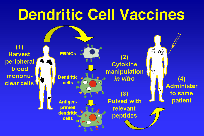
Notwithstanding a decade of use, there still remains a great degree of variability in the development and administration of dendritic cell vaccines. Only a few studies have systematically examined these differences, resulting in a lack of data regarding the most effective means through which to carry out DC-based immunotherapy for CNS neoplasms. Some of these specifics have been resolved on account of information obtained from animal models or through empirical evidence gleaned from common practice. For example, although there are several different methods through which dendritic cells may be acquired, in all of the clinical trials involving DC immunotherapy for glioma to date, they were exclusively manufactured through the differentiation of peripheral blood mononuclear cells (PBMCs) ex vivo. There now exist many methods through which DCs can be produced efficiently and in large enough quantities for clinical trials [5356].

Similarly, no studies exist comparing the efficacy of different sources of antigens in propagating anti-tumor immunity in human patients. The vast repertoire of antigen-loading strategies includes whole tumor cells [27,35,44], apoptotic tumor cells[45], acid-eluted tumor peptides [19,3234], synthetic peptides [20,21], tumor lysate [1417,2126,38,39,43,48], and tumor cDNA/RNA [28,36,40,57]. The effectiveness of these methods in stimulating DC-mediated antitumor immunity has primarily been studied in animal models for proof-of-principle rather than comparative analyses. Although some animal studies have evaluated the efficacy of different sources of antigens in stimulating a DC-mediated anti-tumor response [21,28], the choice of antigenic stimuli in clinical trials seem largely based on previous work with pre-clinical models and theoretical considerations. Clearly, prior experience with a particular DC vaccination protocol allows for ready transition from bench to bedside. Theoretically, however, methods utilizing a wide range of autologous tumor antigens have been favored over peptide selection. This allows the vaccine to target all tumor associated antigens without requisite characterization, helping avoid clonal selection of antigen-loss variants and subsequent tumor-escape [58]. Choice of antigen must also be considered for pragmatic reasons, as poor availability of resected tumor tissue may favor the use of cDNA/RNA to pulse DCs as these antigens are readily amplified through molecular techniques [9]. Still, as most of the antigen sources available to immunotherapy have been shown to prime dendritic cells appropriately, their use may remain largely an empiric choice until future studies comparatively examining their functionality and practicality are conducted.

It is well accepted that antigen loading is most effective when pulsing phenotypically immature dendritic cells. However, the maturation state in which to administer DCs to patients following this step remains unclear. Numerous studies have shown that dendritic cell maturation is necessary for effective DC migration [59] and T-cell stimulation [42,60], thus making them more effective in generating an anti-tumor response [15]. Given the need for an inflammatory stimulus or cytokine to induce dendritic cell maturation, DCs have been matured ex vivo in order to theoretically ensure proper functioning once they reach the lymph nodes of the host [1417]. This is supported by work by Yamanaka et al. who found that patients receiving mature DCs experienced a greater overall survival than patients receiving immature ones [15]. However, Barratt-Boyes et al. were able to demonstrate that immature antigen-pulsed dendritic cells undergo natural maturation when injected intradermally and are quite capable of stimulating appropriate anti-tumor T-cell pathways in vivo. Moreover, they argue that the administration of immature DCs may even be superior to that of mature DCs, as the latter have relatively decreased emigration rates from the injection site [18]. As clinical trials have demonstrated clinical benefit with DC regimens using both immature and mature dendritic cells, future studies comparing the two preparations will be needed to further evaluate the effect maturation status has on clinical efficacy and patient survival [61].

The frequency of DC injections in clinical trials was initially modeled after administration schedules found to be effective in immunotherapy for non-CNS tumors [19]. Since then, the majority of studies have roughly followed a biweekly injection regimen, with number of vaccinations varying from 1 to 22 times (Table 1). Given the many differences in other aspects of the vaccination protocol, it is difficult to compare the efficacy of DC administration frequency between published studies. It has been argued that vaccination should be given expediently following maximal surgical cytoreduction, chemotherapy, and/or radiotherapy in order to fully benefit from the rebound in immune function following gross total resection prior to tumor recurrence [52]. Although early initiation of DC immunotherapy is encouraged, data from animal [26] and patient [14] studies suggest early follow-up vaccinations are not as critical, and in fact may hinder the immune response by causing activation-induced death of recently activated T cells. Instead, these studies have demonstrated that booster injections with tumor lysate alone may be more beneficial in stimulating an anti-tumor response. Interestingly, many studies have employed the testing of delayed type hypersensitivity (DTH) using tumor lysate [1417,37,48], which may have inadvertently served as a form of booster and improved anti-tumor immunity. Given the lack of controlled studies addressing these issues, the timing and frequency of DC vaccine administration remains largely based on empirical experience, and will require future studies to determine an optimal schedule.

The optimal dose of dendritic cells has similarly been questioned. Even from early pre-clinical studies, it was evident that low inoculations of DCs could stimulate an anti-tumor response [16]. Dose escalation protocols in clinical trials have substantiated the finding that DC-mediated immunity is an “on/off” rather than a dose-response phenomenon, as increasing numbers of these APCs do not affect the magnitude of the CTL response [14,33]. This is reassuring, as large quantities of autologous tumor lysate-pulsed dendritic cells were sometimes difficult to obtain during dose escalation protocols [33,40].

Finally, the route of DC administration best for immunotherapy is still under investigation as well. Dendritic cells can be administered through a variety of ways, including subcutaneous, intradermal, intralymphatic, intranodal, and intratumoral injections. Several studies in mice and non-human primates have examined the differences in lymph node accumulation and T-cell stimulation with each route. Radioisotope tracing studies have shown that intravenous DC administration results in the accumulation of dendritic cells within the spleen and liver. However, interestingly this method results in the greatest humoral anti-tumor response as indicated by increased tumor antigen specific antibodies [6265]. Conversely, intradermal [62,65], intralymphatic [62], intracranial [66], intranodal [59], and subcutaneous [63] injections of DCs have been shown to drain to lymph nodes and induce greater T-cell mediated immunity against tumor antigens compared to intravenous injections in pre-clinical models. Much attention has been given to the intranodal or perinodal administration of DCs, as lymph nodes are acknowledged as the processing centers responsible in mediating antigen presentation and T-cell activation [67]. Some investigators have questioned how the placement of these injections may alter the potency of the immune response. Recently Calzascia et al. were able to show that the distance from the cervical nodes was not as critical as the location of the tumor itself [68]. Although there was some improved tissue tropism for the CNS when DCs were administered into cervical lymph nodes, they found that the ultimate determinant of homing signals was the residence of the actual tumor, as was evidenced by CNS-tropic T cells following inguinal node DC injection in an intracranial tumor model. To date, only one clinical trial has investigated the differences in patient outcome between two injection routes. Yamanaka et al. found that patients who received both intratumoral and intradermal DCs had prolonged survival compared to those that received intradermal DCs alone [15]. Further studies comparing injection sites and modalities in inducing antitumor immunity are still needed.

Patient selection for Dendtic Cell Vaccines

The increasing volume of studies reporting on the clinical response to dendritic cell-based immunotherapy has allowed for the analysis of patient demographics to better determine who may benefit the most from this novel treatment modality. As with traditional therapies for malignant brain tumors, younger patients (<40 years old) receiving DC vaccines appeared to do better in terms of overall survival compared to older patients with similar tumor histologies [48,69]. Though this may in part be attributable to the general trend for younger glioma patients to have better prognoses, one particular study was able to demonstrate that this was primarily due to associated declines in thymus function with increasing age [39]. They maintained that CD8+ T-cell production from the thymus was a prognostic indicator of response to DC immunotherapy independent and superseding that of patient age.

Another critical patient characteristic that seems to have an effect on clinical outcome is surgical management. Those patients who underwent gross total resection of their brain tumor experienced significantly better progression free survival (PFS) compared to otherwise similar patients with appreciable residual tumor [14]. Moreover, bulky residual tumor or active tumor recurrence at the time of vaccination appears to debilitate the anti-tumor CTL response [33].

Finally, patients with newly diagnosed malignant glioma seem to achieve greater response rates than those with recurrent tumors [48]. Although these findings remain to be validated by future studies, it would appear that younger patients with newly diagnosed malignant glioma that are amenable to gross total resection would stand to benefit the most from DC based vaccines.

Synergy of Dendritic Cell Vaccines with other therapies

Although much progress has been made in DC-based immunotherapy for CNS tumors, objective clinical responses for vaccinated brain tumor patients remains inconsistent. Consequently, some groups have examined the use of adjuvant treatments to augment the effects of dendritic cell vaccination. These methods include adjuvant chemotherapy, cytokine administration, and toll-like receptor (TLR) agonists.

The use of cytokines to supplement DC-based immunotherapy in human patients is an extension of work done in pre-clinical animal studies [24,27,29]. Although systemic cytokine administration has only been used in one DC vaccine clinical trial to date [44], studies conducted in vitro as well as with human patients have shown that cytokines such as IL-10 [70], IL-18 [71], and IL-23 [72] may enhance the immune response of effector cells in DC immunotherapy. As the data regarding this adjuvant modality is scarce, further studies are needed before routine clinical use of systemic cytokines can be considered.

The use of standard treatments such as chemotherapy to aid immunotherapy has been considered as well. Chemotherapy has traditionally been regarded as an antagonist to the treatment effects of immunotherapy, because of its effects of bone marrow suppression causing lymphopenia. There has also been a belief that the dead apoptotic tumor cells would produce immune tolerance, exacerbating the lymphopenic state that results as well. However, mounting evidence argues that these apoptotic tumor cells may provide a rich antigen source for dendritic cells and that prompt DC vaccination following chemotherapy may actually provide greater benefit than delaying treatment [47]. Recently, studies have shown that when chemotherapy is used adjuvantly with DC-based immunotherapy, patients experience prolonged overall survival as well as increased time to disease progression [45,46,48]. As evidence suggests that chemotherapy in the setting of DC immunotherapy may actually be beneficial rather than obstructive, it may be prudent to further investigate multi-modality treatment strategies for the simultaneous treatment of brain tumor patients.

Conclusion

Over the past decade, dendritic cell-based immunotherapy for CNS tumors has progressed from pre-clinical rodent models and safety assessments to Phase I/II clinical trials in over 200 patients, which have produced measurable immunological responses and some prolonged survival rates. However, many questions regarding the methods and molecular mechanisms behind this new treatment option remain unanswered. Results from currently ongoing and future studies will help to elucidate which dendritic cell preparations, treatment protocols, and adjuvant therapeutic regimens will optimize the efficacy of DC vaccination. Additionally, it will be important to characterize the pathways underlying the immunosuppressive microenvironment of brain tumors that currently hinder anti-tumor responses. Combined with further advances in the manipulation of various lymphocyte subsets such as regulatory T cells and NK-like T cells, in addition to the usual armament of CD4+ & CD8+ T cells, understanding these immunologic intricacies will help maximize the cellular efficiency of immunotherapeutic techniques. As clinical studies continue to report results on DC-mediated immunotherapy, it will be critical to continue refining treatment methods and developing new ways to augment this promising form of glioma treatment.

 
----------------------------------------------------------------------------------------------------------------------------------------------------------


Heat Shock Protein (HSP) is a chaperone protein that helps form proteins, stabilize cells against heat stress and aids in the degradation of proteins. HSP is expressed in response to a rise in body temperature or stress levels elicited by such as infection, inflammation or injury. HSP plays a strong role in antigen presentation, which increase the efficacy of vaccines, including anti-cancer vaccines.

Hsp90, a type of HSP, can stabilize proteins required for tumor growth, which is why Hsp90 inhibitors are being investigated as anti-cancer drugs. By inhibiting Hsp90, the tumor cells become unstable and begin to die. Clinical trials of an anti-cancer vaccine are underway and many in the scientific community hope a viable anti-cancer vaccine will become a reality.

Sources and Research:

Heat shock proteins (HSPs) based anti-cancer vaccines.


STEP ONE

Be informed.  Read information below.

STEP TWO

Schedule consultation with R. Douglas Wichman, MD.

STEP THREE

Submit "New Patient Form", current labwork, & imaging results prior to consultation.

STEP FOUR

Establish if you are a candidate for this therapy

in accordance with the Georgia

"Access to Medical Treatment Act".


Cytokines activate cancer fighting T-Cells within the body

Cytokine Therapy Benefits

  • Uses the body’s immune system to fight cancer.
  • Activates T-Cells, which destroy cancer cells.
  • Prevents cancer spread.
  • Enhances the healing of normal cells damaged by chemotherapy or radiation therapy.

In the fight against cancer, one of the first steps is boosting the immune system to nhance the body’s natural ability to fight invaders. Immunotherapy has proven successful in decreasing cancer cells in the body. An integral part of immunotherapy is the administering of cytokines. Cytokines are types of molecules that are secreted by cells which communicate with other cells. Typical cytokines are interferon or interleukin. However, some cytokines can either help or hinder cancer growth. By using the right cytokine type, it can communicate with cancer fighting T-cells and stimulate macrophages, cells that consume toxic substances

Using Cytokines to Fight Cancer:

Cytokine Cancer Therapy uses either natural or lab produced cytokines to help the immune system react to cancer cells and destroy them. Cytokine Therapy seeks out cancer cells throughout the body and also prevents growth. Cytokines are also known as biological response modifiers, which change how the immune system interacts with cancer cells. A typical therapy involves adding interferons or interleukins into the bloodstream. Both of these cytokines communicate with the immune system and facilitate type 1 helper T cells, which are the key to fighting cancer cells. T helper cells are an aggressive type of white blood cell which can activate cancer cell killing T cells and enhance activity of macrophages – cells that consume malignant substances. Now that T cells are activated, the immune system fights the cancer cells and can reduce its growth and in some cases can diminish the cancer mass and reduce its threat. Cytokine Therapy is used along with other cancer treatments and can assist in healing cell damage from chemotherapy or radiation therapy.

Hyperbaric Oxygen Therapy


Hyperbaric Oxygen Therapy (HBOT) helps all kinds of people. It’s used to help patients suffering from sports injuries, Chronic Fatigue Syndrome, infections, arthritis and a huge range of other medical conditions. As we all know, oxygen is vital for life, and life cannot exist without it. It follows that oxygen is essential for effective healing and recovery.

read more

Book Your Appointment NOW

Call (770) 232-7883


Click to E-Mail Us Now!


Our Address:

4488 N. Shallowford Road, Suite 201

Dunwoody, Georgia 30338


We Serve the Following Areas:

Atlanta, Georgia, Acworth, Alpharetta, Berkeley Lake, Braselton, Brookhaven, Buckhead, Buford, Canton, Chamblee, Conyers, Cumming, Dacula, Decatur, Doraville, Duluth, Dunwoody, Georgia, Flowery Branch, Gainesville, Grayson, Hoschton, Johns Creek, Kennesaw, Lawrenceville, Lilburn, Lithonia, Loganville, Marietta, Milton, Norcross, Roswell, Sandy Springs, Smyrna, Snellville, Stone Mountain, Sugar Hill, Suwanee, Tucker, Vinings, Woodstock, Georgia